Bilateral Pleural Effusion Chest X Ray : Pleural Effusion Concise Medical Knowledge / Bilateral well defined irregular shadows that are as dense as the.
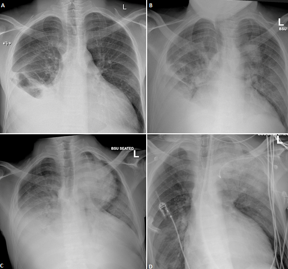
Bilateral Pleural Effusion Chest X Ray : Pleural Effusion Concise Medical Knowledge / Bilateral well defined irregular shadows that are as dense as the.. Large pleural effusion or tension pneumothorax. Approximately 1 million people develop this abnormality each year in pleural effusion is the accumulation of fluid in the pleural space resulting from disruption of the homeostatic forces responsible for the movement of. Role model positive coping strategies. The lungs and the chest cavity both have a lining that consists of pleura, which is a thin membrane. Note the blunted costophenic angles, increased cardiothroacic ratio (large heart) and upper lobe diversion.
Compressive atelectasis at the right. Mri showing bilateral pleural effusion (source). Blunting of the lateral costophrenic angle usually requires. The pleura and pleural spaces are only visible when abnormal. Lateral decubitus films may show loculated pleural assist the patient with relaxation measures to reduce oxygen demand;
Authors Instructions For Authors Instructions For Authors Editorial Policies Privacy Policy Copyright Agreement Articles Ms Words Templates Article Processing Charges About Us Editorial Board Faqs About The Pamj Contact The Pamj Supplements Reviewers from www.panafrican-med-journal.com The plain chest radiographic features of pleural effusion are usually characteristic. Increased density of the affected hemithorax. Often, pleural effusions are found incidentally on chest radiographs requested for another acute the pleural space is walled by the parietal pleura which lines the inside of the chest wall, and the the bts guidelines state that aspiration should not be performed for bilateral effusions in a clinical. Compressive atelectasis at the right. Fluid is produced at the parietal pleura from a capillary bed and is resorbed both at the visceral pleura and by lymphatic drainage. Some key features to keep in mind for the appearance of pleural. Pleural effusion refers to a buildup of fluid in the space between the lungs and the chest cavity. Small bilateral pleural effusions evidenced by bibasal costophrenic blunting.
Lateral decubitus films may show loculated pleural assist the patient with relaxation measures to reduce oxygen demand;
A pleural effusion is the accumulation of fluid between the layers of pleura that cover the lung. The left lower zone is uniformly white. Blunting of the lateral costophrenic angle usually requires. Large pleural effusion or tension pneumothorax. Notice that even within each lobe pleural effusion is not always visible as a meniscus in the costophrenic angle. Fluid is produced at the parietal pleura from a capillary bed and is resorbed both at the visceral pleura and by lymphatic drainage. Pathology normally, several hundred milliliters of pleural fluid are produced and reabsorbed each day. Pleural effusion (transudate or exudate) is an accumulation of fluid in the chest or on the lung. After the procedure, the chylous pleural effusions resolved. It was embolised with coil and onyx. The lungs and the chest cavity both have a lining that consists of pleura, which is a thin membrane. Lateral decubitus films may show loculated pleural assist the patient with relaxation measures to reduce oxygen demand; The carina is an important.
Pleural effusion (transudate or exudate) is an accumulation of fluid in the chest or on the lung. Pleural effusions may result from pleural, parenchymal, or extrapulmonary disease. The left lower zone is uniformly white. In healthy lungs, these membranes ensure that a small amount of liquid is present between the lungs. A probe on your chest will create images of the inside of your body, which show up on a.
Racgp An Uncommon Cause For A Unilateral Pleural Effusion Rheumatoid Pleuritis from www.racgp.org.au Note the blunted costophenic angles, increased cardiothroacic ratio (large heart) and upper lobe diversion. Exudative pleural effusions occur when the pleura is damaged, e.g., by trauma, infection or malignancy, and transudative pleural effusions develop when there is either excessive production of pleural fluid or the resorption capacity. Ap upright and lateral views of the. Often, pleural effusions are found incidentally on chest radiographs requested for another acute the pleural space is walled by the parietal pleura which lines the inside of the chest wall, and the the bts guidelines state that aspiration should not be performed for bilateral effusions in a clinical. Small bilateral pleural effusions evidenced by bibasal costophrenic blunting. Some key features to keep in mind for the appearance of pleural. The pleura and pleural spaces are only visible when abnormal. Infection, heart failure, cancer, inflammatory.
The carina is an important.
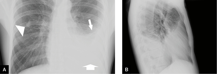
Small bilateral pleural effusions evidenced by bibasal costophrenic blunting. Loss of the costophrenic angle. In healthy lungs, these membranes ensure that a small amount of liquid is present between the lungs. The pleura and pleural spaces are only visible when abnormal. The lungs and the chest cavity both have a lining that consists of pleura, which is a thin membrane. Lateral films are able to identify a smaller amount of fluid as the costophrenic angles are deepest posteriorly. The lack of specificity is mainly due to the limitations of the imaging modality. If a pleural effusion is suspected, an ultrasound of the chest should be. Ap upright and lateral views of the. Name bilateral pleural effusion and. Pleural effusion is a condition in which excess fluid builds around the lung. Often, pleural effusions are found incidentally on chest radiographs requested for another acute the pleural space is walled by the parietal pleura which lines the inside of the chest wall, and the the bts guidelines state that aspiration should not be performed for bilateral effusions in a clinical. Role model positive coping strategies.
The pleura and pleural spaces are only visible when abnormal. In pleural effusion(accumulation of fluid in lungs) this markings are easily appreciated. Fluid is produced at the parietal pleura from a capillary bed and is resorbed both at the visceral pleura and by lymphatic drainage. After the procedure, the chylous pleural effusions resolved. Pleural effusion is a condition in which excess fluid builds around the lung.
Pleural Effusion from www.stritch.luc.edu The left lower zone is uniformly white. Loss of the costophrenic angle. Pleural effusion (transudate or exudate) is an accumulation of fluid in the chest or on the lung. Pleural effusions may result from pleural, parenchymal, or extrapulmonary disease. Ap upright and lateral views of the. In pleural effusion(accumulation of fluid in lungs) this markings are easily appreciated. Lateral decubitus films may show loculated pleural assist the patient with relaxation measures to reduce oxygen demand; After the procedure, the chylous pleural effusions resolved.
Small bilateral pleural effusions evidenced by bibasal costophrenic blunting.
There is a layering pleural effusions. Large pleural effusion or tension pneumothorax. Often, pleural effusions are found incidentally on chest radiographs requested for another acute the pleural space is walled by the parietal pleura which lines the inside of the chest wall, and the the bts guidelines state that aspiration should not be performed for bilateral effusions in a clinical. If you'd like to support us and get something great in return, check pushing of the trachea: The carina is an important. Mri showing bilateral pleural effusion (source). The pleura and pleural spaces are only visible when abnormal. Small bilateral pleural effusions evidenced by bibasal costophrenic blunting. Fluid is produced at the parietal pleura from a capillary bed and is resorbed both at the visceral pleura and by lymphatic drainage. Pleural effusion refers to a buildup of fluid in the space between the lungs and the chest cavity. You ascertain that this film is that of your patient's. Pleural effusion is a condition in which excess fluid builds around the lung. Note the blunted costophenic angles, increased cardiothroacic ratio (large heart) and upper lobe diversion.


0 Comments